Login
Registration enables users to use special features of this website, such as past
order histories, retained contact details for faster checkout, review submissions, and special promotions.
order histories, retained contact details for faster checkout, review submissions, and special promotions.
Forgot password?
Registration enables users to use special features of this website, such as past
order histories, retained contact details for faster checkout, review submissions, and special promotions.
order histories, retained contact details for faster checkout, review submissions, and special promotions.
Quick Order
Products
Antibodies
ELISA and Assay Kits
Research Areas
Infectious Disease
Resources
Purchasing
Reference Material
Contact Us
Location
Corporate Headquarters
Vector Laboratories, Inc.
6737 Mowry Ave
Newark, CA 94560
United States
Telephone Numbers
Customer Service: (800) 227-6666 / (650) 697-3600
Contact Us
Additional Contact Details
Login
Registration enables users to use special features of this website, such as past
order histories, retained contact details for faster checkout, review submissions, and special promotions.
order histories, retained contact details for faster checkout, review submissions, and special promotions.
Forgot password?
Registration enables users to use special features of this website, such as past
order histories, retained contact details for faster checkout, review submissions, and special promotions.
order histories, retained contact details for faster checkout, review submissions, and special promotions.
Quick Order
IRS1
insulin receptor substrate 1
May mediate the control of various cellular processes by insulin. When phosphorylated by the insulin receptor binds specifically to various cellular proteins containing SH2 domains such as phosphatidylinositol 3-kinase p85 subunit or GRB2. Activates phosphatidylinositol 3-kinase when bound to the regulatory p85 subunit.
| Gene Name: | insulin receptor substrate 1 |
| Synonyms: | IRS1, IRS-1, HIRS-1, Insulin receptor substrate 1 |
| Target Sequences: | NM_005544 NP_005535.1 P35568 |
Publications (4)
1
Antisense treatment of IGF-IR enhances chemosensitivity in squamous cell carcinomas of the head and neck. Liu S, Jin F, Dai W, Yu Y. European journal of cancer (Oxford, England : 1990). 2010 46:1744-51. (WB)
[PubMed:20451373]
2
The reciprocal regulation of gamma-synuclein and IGF-I receptor expression creates a circuit that modulates IGF-I signaling. Li M, Yin Y, Hua H, Sun X, Luo T, Wang J, Jiang Y. The Journal of biological chemistry. 2010 285:30480-8. (WB)
[PubMed:20670935]
[PMC:PMC2945541]
3
Phospho-?Np63a/microRNA network modulates epigenetic regulatory enzymes in squamous cell carcinomas. Ratovitski EA. Cell cycle (Georgetown, Tex.). 2014 13:749-61. (WB; Human)
[Full Text Article]
[PubMed:24394434]
[PMC:PMC3979911]
☰ Filters
Products
Antibodies
(152)
ELISA Kits
(18)
Proteins
(5)
Type
Cell-Based
(1)
Cell-Based Phosphorylation
(7)
Custom
(2)
Over-Expression Cell Culture Supernatant
(1)
Over-Expression Lysate
(3)
Primary
(152)
Recombinant
(1)
Sandwich
(8)
Target
IRS1
(175)
Reactivity
Human
(146)
Mouse
(133)
Rat
(125)
Bovine
(7)
Chicken
(3)
Dog
(8)
Horse
(2)
Monkey
(21)
Pig
(11)
Rabbit
(2)
Xenopus
(1)
Application
IHC
(91)
IHC-P
(37)
WB
(124)
ELISA
(37)
ICC
(26)
IF
(57)
IP
(28)
Peptide-ELISA
(22)
Host
rabbit
(123)
mouse
(29)
Product Group
IHCPlus
(2)
Isotype
IgG
(82)
IgG1
(8)
IgG1,k
(6)
IgG2a
(2)
IgG2a,k
(12)
Clonality
monoclonal mc
(29)
polyclonal pc
(123)
Clone
D51C3
(1)
OTI1D10
(2)
OTI2B10
(2)
OTI3G10
(2)
OTI9E11
(2)
TS106
(1)
Format
12 x 8-Well Microstrips
(1)
96-Well Microplate
(9)
96-Well Strip Plate
(8)
APC
(1)
APC Conjugated
(4)
Biotin Conjugated
(5)
Carrier-free
(4)
Cy3 Conjugated
(1)
Cy7 Conjugated
(1)
FITC Conjugated
(4)
HRP Conjugated
(4)
PE Conjugated
(4)
Unconjugated
(129)
Epitope
pSer307
(13)
aa1-280
(8)
aa837-1089
(8)
pSer312
(7)
Mouse pSer307 and Human pSer312
(6)
pSer636
(6)
Internal
(5)
Mouse pSer302 and Human pSer307
(5)
pSer639
(4)
250-330 aa
(3)
pSer1101
(3)
pSer323
(3)
pSer794
(3)
570-650 aa
(2)
Arg301
(2)
Tyr896
(2)
aa1192-1242
(2)
aa1222-1235
(2)
aa801-1020
(2)
pSer612
(2)
1040-1120 aa
(1)
260-340 aa
(1)
550-630 aa
(1)
580-660 aa
(1)
730-810 aa
(1)
840-920 aa
(1)
Ab-612
(1)
Ab-794
(1)
Asp606
(1)
C-Terminus
(1)
Glu306
(1)
Gly630
(1)
Leu789
(1)
Pro618
(1)
Ser1101
(1)
Ser318
(1)
Ser616
(1)
Ser636
(1)
Ser639
(1)
Tyr632
(1)
Val317
(1)
aa1-160
(1)
aa1041-1242
(1)
aa106-122
(1)
aa1067-1116
(1)
aa274-323
(1)
aa279-328
(1)
aa289-338
(1)
aa431-446
(1)
aa578-627
(1)
aa603-652
(1)
aa605-654
(1)
aa750-800
(1)
aa760-809
(1)
p307
(1)
p312
(1)
p636
(1)
p639
(1)
pSer302
(1)
pSer318
(1)
pSer616
(1)
pSer789
(1)
pTyr896
(1)
Publications
No
(174)
Yes
(1)
Sample Type
Adherent Cell Cultures
(8)
Cell Culture Supernatants
(1)
Cell Lysates
(3)
Plasma
(5)
Serum
(5)
Tissue Homogenates
(5)
Tag
6His, N-terminus
(2)
His-T7
(1)
Myc-DDK (Flag)
(2)
Species
Human
(4)
Mouse
(1)
Source
293T Cells
(1)
E. coli
(1)
HEK 293 Cells
(3)
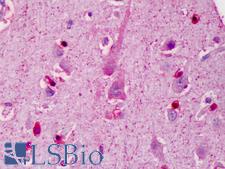
IRS1 Rabbit anti-Human Polyclonal Antibody
Mouse, Human
ELISA, ICC, IF, IHC, IHC-P, WB
Unconjugated
0.05 mg/$375
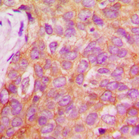
IRS1 Rabbit anti-Human Polyclonal (pSer1101) Antibody
Mouse, Rat, Pig, Human, Monkey
IHC, IHC-P, WB
Unconjugated
30 µl/$263; 50 µl/$279; 100 µl/$315; 200 µl/$379
Sandwich
96-Well Strip Plate
Rat
0.312 - 20 ng/ml
Colorimetric - 450nm (TMB)
Plasma, Serum
1 Plate/$669
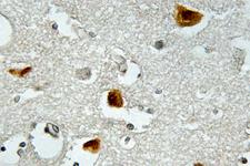
IRS1 Rabbit anti-Human Polyclonal (pSer636) Antibody
Mouse, Rat, Human
IF, IHC, IHC-P, WB
Unconjugated
50 µl/$328; 100 µl/$419
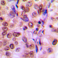
IRS1 Rabbit anti-Human Polyclonal (Internal) Antibody
Mouse, Rat, Pig, Human
ICC, IF, IHC, IHC-P, WB
Unconjugated
30 µl/$255; 50 µl/$271; 100 µl/$299; 200 µl/$351
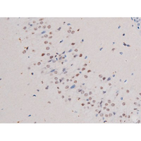
IRS1 Rabbit anti-Human Polyclonal (pSer636) Antibody
Mouse, Rat, Human, Monkey
IF, IHC, Peptide-ELISA, WB
Unconjugated
100 µl/$282; 200 µl/$350
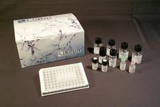
Sandwich
96-Well Strip Plate
Human
0.313 - 20 ng/ml
Colorimetric - 450nm (TMB)
Plasma, Serum, Tissue Homogenates
1 Plate/$579
IRS1 Rabbit anti-Mouse Monoclonal (pSer318) (D51C3) Antibody
Mouse, Rat, Human
IP, WB
Unconjugated
100 µl/$689
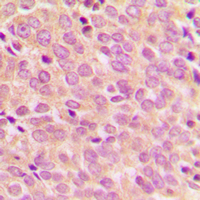
IRS1 Rabbit anti-Human Polyclonal (pSer794) Antibody
Chicken, Rat, Human
ICC, IF, IHC, IHC-P, WB
Unconjugated
30 µl/$263; 50 µl/$279; 100 µl/$315; 200 µl/$379
IRS1 Mouse anti-Mouse Monoclonal (pSer302) Antibody
Mouse, Rat, Human
IF, IHC, IP, WB
Unconjugated
100 µg/$880
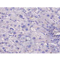
IRS1 Rabbit anti-Human Polyclonal (pSer307) Antibody
Mouse, Rat, Human, Monkey
IF, IHC, Peptide-ELISA, WB
Unconjugated
100 µl/$282; 200 µl/$350
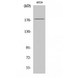
IRS1 Rabbit anti-Human Polyclonal (570-650 aa) Antibody
Mouse, Rat, Human
ELISA, IF, IHC, WB
Unconjugated
50 µg/$295; 100 µg/$335; 200 µg/$394
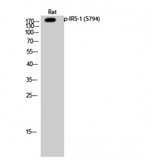
IRS1 Rabbit anti-Human Polyclonal (730-810 aa) Antibody
Mouse, Rat, Human
ELISA, IF, IHC, WB
Unconjugated
50 µg/$295; 100 µg/$335; 200 µg/$394
Sandwich
96-Well Strip Plate
Human
0.313 - 20 ng/ml
Colorimetric - 450nm (TMB)
Cell Lysates, Tissue Homogenates
1 Plate/$730
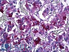
IRS1 Rabbit anti-Human Polyclonal (aa289-338) Antibody
Mouse, Rat, Human
IF, IHC, IHC-P, Peptide-ELISA
Unconjugated
50 µl/$375
![IRS1 Antibody - Anti-IRS1 pS307 Antibody - Western Blot. Western blot of Affinity Purified anti-IRS1 pS307 antibody shows detection of a band at ~180 kD believed to represent phosphorylated IRS1 (arrowhead). Lane 1 shows staining of human 293 cell lysate. Lane 2 shows staining of 293 cell lysate prepared from cells serum-starved for 18 h followed by treatment with 5 ug/ml of anisomysin for 30 min. The pronounced staining of the band at 180 kD is not seen when the antibody was pre-incubated with immunizing peptide prior to reaction (data not shown). The identity of the intensely reactive bands at ~70 kD in both lane 1 and 2 is unknown, although these bands were also competed out by pre-incubation with the immunizing peptide. Approximately 25 ug of each lysate was separated on a 4-20% Tris-Glycine gel by SDS-PAGE and transferred onto nitrocellulose. After blocking with 5% goat serum, 0.5% BLOTTO in PBS, the membrane was probed with the primary antibody diluted to 1:250. Reaction occurred overnight at 4C followed by washes and reaction with a 1:10000 dilution of IRDye800 conjugated Gt-a-Rabbit IgG [H&L] MX ( for 45 min at room temperature (800 nm channel, green). Molecular weight estimation was made by comparison to prestained MW markers in lane M (700 nm channel, red). IRDye800 fluorescence image was captured using the Odyssey Infrared Imaging System developed by LI-COR. IRDye is a trademark of LI-COR, Inc. Other detection systems will yield similar results.](https://lsbio-7d62.kxcdn.com/image2/irs1-antibody-phospho-ser307-ls-c18824/62832_4909668.jpg)
IRS1 Rabbit anti-Human Polyclonal (pSer307) Antibody
Human
ELISA, IHC, WB
Unconjugated
100 µg/$567
IRS1 Mouse anti-Human Monoclonal (aa431-446) Antibody
Mouse, Dog, Bovine, Rat, Pig, Human, Monkey
ICC, IP, WB
Unconjugated
100 µg/$880
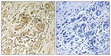
IRS1 Rabbit anti-Human Polyclonal (pSer639) Antibody
Mouse, Rat, Human
IHC, IHC-P, Peptide-ELISA
Unconjugated
50 µl/$334; 100 µl/$397
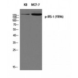
IRS1 Rabbit anti-Human Polyclonal (840-920 aa) Antibody
Mouse, Rat, Human
ELISA, WB
Unconjugated
50 µg/$295; 100 µg/$335; 200 µg/$394
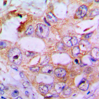
IRS1 Rabbit anti-Human Polyclonal (pSer636) Antibody
Mouse, Rat, Pig, Human
ICC, IF, IHC, IHC-P, WB
Unconjugated
30 µl/$263; 50 µl/$279; 100 µl/$315; 200 µl/$379
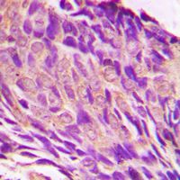
IRS1 Rabbit anti-Human Polyclonal (pSer307) Antibody
Mouse, Rat, Human, Monkey
ICC, IF, IHC, IHC-P, WB
Unconjugated
30 µl/$263; 50 µl/$279; 100 µl/$315; 200 µl/$379
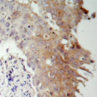
IRS1 Rabbit anti-Human Polyclonal (Internal) Antibody
Mouse, Rat, Human
ICC, IF, IHC, IHC-P, WB
Unconjugated
30 µl/$266; 50 µl/$286; 100 µl/$321; 200 µl/$386
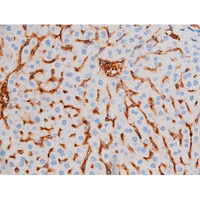
IRS1 Rabbit anti-Human Polyclonal (pSer312) Antibody
Mouse, Rat, Human, Monkey
IF, IHC, Peptide-ELISA, WB
Unconjugated
100 µl/$282; 200 µl/$350
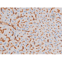
IRS1 Rabbit anti-Human Polyclonal (pSer639) Antibody
Mouse, Rat, Human, Monkey
IF, IHC, Peptide-ELISA, WB
Unconjugated
100 µl/$282; 200 µl/$350
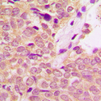
IRS1 Rabbit anti-Human Polyclonal (Internal) Antibody
Mouse, Rat, Human, Monkey
ICC, IF, IHC, IHC-P, WB
Unconjugated
30 µl/$255; 50 µl/$271; 100 µl/$299; 200 µl/$351
Viewing 1-25
of 175
product results
If you do not find the reagent or information you require, please contact Customer.Support@LSBio.com to inquire about additional products in development.


