Primary IHC Markers In Breast Cancer
Login
Registration enables users to use special features of this website, such as past
order histories, retained contact details for faster checkout, review submissions, and special promotions.
order histories, retained contact details for faster checkout, review submissions, and special promotions.
Forgot password?
Registration enables users to use special features of this website, such as past
order histories, retained contact details for faster checkout, review submissions, and special promotions.
order histories, retained contact details for faster checkout, review submissions, and special promotions.
Quick Order
Products
Antibodies
ELISA and Assay Kits
Research Areas
Infectious Disease
Resources
Purchasing
Reference Material
Contact Us
Location
Corporate Headquarters
Vector Laboratories, Inc.
6737 Mowry Ave
Newark, CA 94560
United States
Telephone Numbers
Customer Service: (800) 227-6666 / (650) 697-3600
Contact Us
Additional Contact Details
Login
Registration enables users to use special features of this website, such as past
order histories, retained contact details for faster checkout, review submissions, and special promotions.
order histories, retained contact details for faster checkout, review submissions, and special promotions.
Forgot password?
Registration enables users to use special features of this website, such as past
order histories, retained contact details for faster checkout, review submissions, and special promotions.
order histories, retained contact details for faster checkout, review submissions, and special promotions.
Quick Order
Breast Cancer Immunohistochemistry Markers
IHC markers are used in both clinical and research settings to provide insights into a variety of processes that are of interest to breast cancer and disease research at large.
Breast cancer, the most common malignancy in women worldwide, is classified by using microscopic morphologic criteria along with standard clinical immunohistochemistry markers
for HER2 and ER/PR
in order to predict if a patient’s tumor will respond to Herceptin or hormone therapy. IHC is further useful for determining rates of cell differentiation, elucidating molecular pathways, and
highlighting the proteomics of tumor growth and metastatic potential. For example, IHC markers help distinguish in-situ from invasive carcinomas (KRT14
and KRT5), subtype ductal from lobular carcinomas (E-cadherin,
p120, FOXA1, GATA3),
determine mammary origin of a metastatic carcinoma (CK7, CK20,
Mammaglobin A), and highlight tumor proliferation and apoptosis (Ki-67,
BCL2, TP53). Furthermore, the myoepithelial markers
SMA (Smooth Muscle Actin), Calponin,
p63 and SMMHC can be used to determine whether or not a cancer has invaded, since
benign and early lesions have an intact myoepithelial layer surrounding breast glands.
Targeting pathways through IHC:
IHC markers can be used to identify the deregulation or pathogenic activation of various cell-cycle pathways in breast cancer. These include BCL2,
an anti-apoptotic protein which promotes...
Click to Expand Text ▸
ERBB2 / HER2
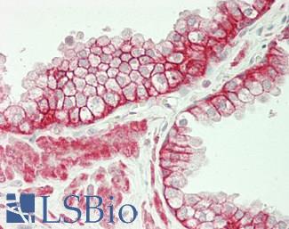
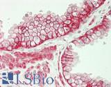
HER2 (ERBB2) is a tyrosine kinase/ epidermal growth factor receptor that is overexpressed and mutated in approximately 10-20% of breast cancers (Zaha, 2014). ERBB2 (HER2) exhibits positive membranous staining in malignant cells, where it promotes cancer cell growth. It is associated with more aggressive tumors. Immunohistochemistry staining of ERBB2 is used alongside the proteins ER (estrogen receptor), PR (progesterone receptor) and Ki-67 to classify different subtypes of breast cancer, and it is a widely used prognostic marker for breast cancer, as levels of ERBB2 expression predict a patient’s response to Herceptin therapy.
Staining:
While this target can show either cytoplasmic or membranous staining, only membranous staining is relevant for cancer.
LSBio's recommended antibody to HER2 for use in immunohistochemistry is HER2 Antibody LS‑B2133.
Click here for specifications and further information on this antibody.
Click here for specifications and further information on this antibody.
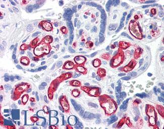
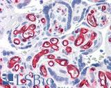
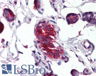
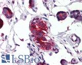
 Anti-ERBB2 / HER2 antibody IHC of human breast, carcinoma. Immunohistochemistry of formalin-fixed, paraffin-embedded tissue after heat-induced antigen retrieval. Antibody LS-B2133 concentration 5 ug/ml.
HER2 Antibody LS-B2133
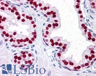
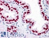
Estrogen Receptor / ESR1
ER (Estrogen receptor / ESR1) is a biomarker that is prognostic for breast cancer and predicts response to hormonal therapy. Estrogen receptor is a nuclear hormone receptor with nuclear staining in malignant breast cancer cells, and usually has higher levels of expression in Luminal A subtypes compared to others (Jeselsohn, 2015). Levels of expression of estrogen receptor are used in conjunction with progesterone receptor (PR), HER2 and Ki-67 expression levels to subtype breast cancers for different downstream treatments. Most breast cancers have positive expression of ER protein, primarily in luminal tissues where the luminal A subtype sees high levels of expression (Jeselsohn, 2015; Yersal, 2014).
Staining:
Staining for this target is expected to be nuclear.
LSBio's recommended antibody to ER for use in immunohistochemistry is ER Antibody LS‑B10527.
Click here for specifications and further information on this antibody.
Click here for specifications and further information on this antibody.
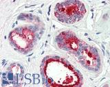
 Anti-ER Alpha / Estrogen Receptor antibody IHC staining of human breast. Immunohistochemistry of formalin-fixed, paraffin-embedded tissue after heat-induced antigen retrieval. Antibody LS-B10527 dilution 1:100.
ER Antibody LS-B10527
Progesterone Receptor / PGR
PR (progesterone receptor / PGR) is a biomarker that is prognostic for breast cancer and predicts response to hormonal therapy. Progesterone receptor is a nuclear hormone with nuclear staining in malignant breast cancer cells. Immunohistochemistry staining of PR is used alongside estrogen receptor (ER), HER2 and Ki-67 expression levels to subtype breast cancers for different downstream treatments. PR has been found to modulate the behavior of estrogen receptor protein and impede the growth of ER positive tumors, which coincides with the typically better prognosis of ER+, PR+ breast cancers when compared to other subtypes (Mohammed, 2015).
Staining:
Staining for this target is expected to be nuclear.
LSBio's recommended antibody to PR for use in immunohistochemistry is PR Antibody LS‑B2983.
Click here for specifications and further information on this antibody.
Click here for specifications and further information on this antibody.
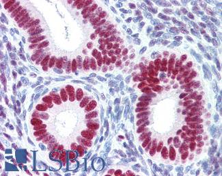
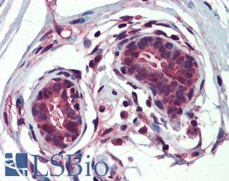
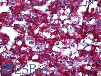
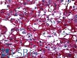
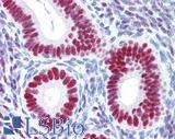 Anti-PGR / Progesterone Receptor antibody IHC of human uterus. Immunohistochemistry of formalin-fixed, paraffin-embedded tissue after heat-induced antigen retrieval. Antibody LS-B2983 concentration 20 ug/ml.
PR Antibody LS-B2983
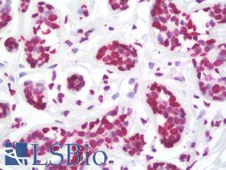
TP53
TP53 is a tumor suppressor that may be used as a marker for estrogen and progesterone receptor negative high grade breast cancers. It is associated with basal subtypes, where it may have aberrant or positive expression (Zaha, 2014). High levels of TP53 protein are often found in triple negative breast cancers (TNBC) alongside high levels of Ki-67. In TNBC, TP53 staining correlates with tumor grade and worse prognosis (Pan, 2017).
Staining:
TP53 staining is typically nuclear, but antibodies to this target can also show weaker cytoplasmic staining.
LSBio's recommended antibody to TP53 for use in immunohistochemistry is TP53 Antibody LS‑C172956.
Click here for specifications and further information on this antibody.
Click here for specifications and further information on this antibody.

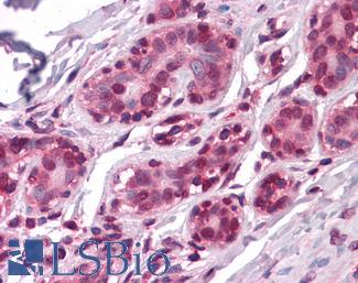
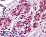
 IHC of paraffin-embedded Carcinoma of Human lung tissue using anti-TP53 mouse monoclonal antibody.
TP53 Antibody LS-C172956
EGFR
EGFR protein regulates a number of fundamental cell cycle pathways and is involved in migration and tumor invasion. Gene amplification of EGFR is a frequent occurrence in some types of breast cancer and also in numerous other tissues. Roughly half of triple negative and also IBC (inflammatory breast cancer) cases show overexpression of this protein (Masuda, 2012). Expression is generally associated with the basal subtype but is also seen in some luminal B tumors, and is correlated with lower ER / PR expression (Changavi, 2015).
Staining:
This protein has membranous and cytoplasmic staining in IHC.
LSBio's recommended antibody to EGFR for use in immunohistochemistry is EGFR Antibody LS‑B2914.
Click here for specifications and further information on this antibody.
Click here for specifications and further information on this antibody.
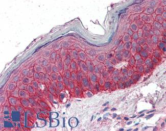
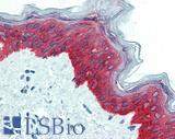
 Anti-EGFR antibody IHC of human skin. Immunohistochemistry of formalin-fixed, paraffin-embedded tissue after heat-induced antigen retrieval. Antibody LS-B2914 concentration 5 ug/ml.
EGFR Antibody LS-B2914
BRCA1
BRCA1 is a multifunctional protein involved in DNA repair, tumor suppression and cell replication, whose dysfunction through inherited or somatic mutation or through epigenetic silencing is the oncogenic source of a certain percentage of breast cancers (Romagnolo, 2015). Mutations in this gene confer high risk for both breast cancer and ovarian cancer, and thus immunohistochemistry staining and testing for wild type and mutated forms of this repair gene are necessary for diagnosis and downstream treatment (Anantha, 2017).
Staining:
This protein has nuclear and cytoplasmic staining in IHC depending upon the isoform.
LSBio's recommended antibody to BRCA1 for use in immunohistochemistry is BRCA1 Antibody LS‑B3772.
Click here for specifications and further information on this antibody.
Click here for specifications and further information on this antibody.
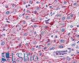
 Anti-BRCA1 antibody IHC of human breast. Immunohistochemistry of formalin-fixed, paraffin-embedded tissue after heat-induced antigen retrieval. Antibody LS-B3772 concentration 5 ug/ml.
BRCA1 Antibody LS-B3772
APOBEC3B
APOBEC3B is a repair gene and cytidine deaminase involved in somatic hypermutation. The deregulation or dysfunction of APOBEC3B and other members of the APOBEC family has been implicated as a direct source of mutagenesis in breast and other cancers (Nik-Zainal, 2012; McBride, 2008; Hwang, 2015). APOBEC3B is upregulated in a number of breast cancers, and this upregulation correlates with a mutation spectrum (kataegis) in these tumors consistent with APOBEC function and may be a simultaneous or downstream consequence of ERBB2 amplification (Nik-Zainal, 2012; Kanu, 2016).
Staining:
Staining for APOBEC3B is expected to be nuclear.
LSBio's recommended antibody to APOBEC3B for use in immunohistochemistry is APOBEC3B Antibody LS‑B12051.
Click here for specifications and further information on this antibody.
Click here for specifications and further information on this antibody.
Proliferation Markers In Breast Cancer
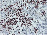
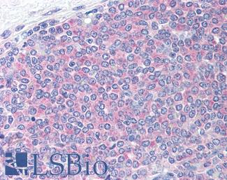
Ki-67 (MKI67)
Ki-67 (MKI67) is a marker for proliferation in breast cancer cells and many other tumor and tissue types. The degree of expression of Ki-67 is used in conjunction with ER, PR and HER2 expression levels to subtype breast cancers. Higher levels of this protein are correlated with increased rates of proliferation (Zaha, 2014), absence of ER and PR and poor prognosis, and staining for Ki-67 in immunohistochemistry is thus important for determining the necessary treatment (Zorka, 2014).
Staining:
Staining for this target is expected to be nuclear.
LSBio's recommended antibody to Ki-67 for use in immunohistochemistry is Ki-67 Antibody LS‑B3321.
Click here for specifications and further information on this antibody.
Click here for specifications and further information on this antibody.
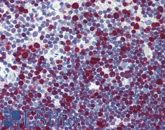
 Anti-MKI67 / Ki-67 antibody IHC of human thymus. Immunohistochemistry of formalin-fixed, paraffin-embedded tissue after heat-induced antigen retrieval. Antibody LS-B3321 dilution 1:50.
Ki-67 Antibody LS-B3321
Cell-Type Specific Markers In Breast Cancer
CNN1 (Calponin)
CNN1 (calponin) is a myoepithelial marker in breast cancer with functional roles in proliferation, angiogenesis and migration (O’Kelly, 2008). In immunohistochemistry staining, calponin is found circulating the nucleus in the cytoplasm of myoepithelial cells (Zaha, 2014).
LSBio's recommended antibody to Calponin for use in immunohistochemistry is Calponin Antibody LS‑B4304.
Click here for specifications and further information on this antibody.
Click here for specifications and further information on this antibody.
 Anti-CNN1 / Calponin antibody IHC of human myometrium. Immunohistochemistry of formalin-fixed, paraffin-embedded tissue after heat-induced antigen retrieval. Antibody LS-B4304 concentration 5 ug/ml.
Calponin Antibody LS-B4304
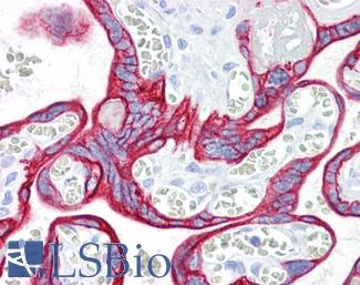
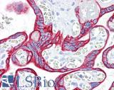
Smooth Muscle Actin (SMA / ACTA2)
Smooth Muscle Actin (SMA / ACTA2) is a myoepithelial marker for normal and cancerous breast tissue. Smooth muscle actin is often used alongside SMMHC, calponin and cytokines like CK5 and CK17 to distinguish myoephithelial cells from ductal carcinoma cells, which are negative for these proteins (Zaha, 2014). High levels of smooth muscle actin expression are correlated with poorer prognosis in breast cancers positive for EGFR and ERBB2; breast cancers with overexpression of these proteins see upregulation of ACTA2 likely due to its regulation by the upstream transcription activator STAT1. Overexpression of ACTA2 is correlated with an increase in invasive potential through its functions in the epithelial to mesenchymal transition (Jeon, 2016).
Staining:
Staining for this target is cytoplasmic.
LSBio's recommended antibody to SMA for use in immunohistochemistry is SMA Antibody LS‑B7351.
Click here for specifications and further information on this antibody.
Click here for specifications and further information on this antibody.
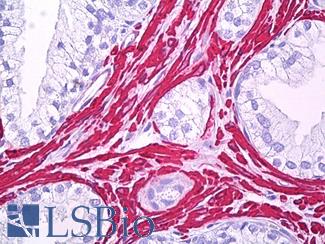
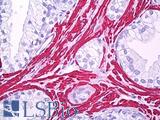 Anti-Smooth Muscle Actin antibody IHC of human prostate, smooth muscle. Immunohistochemistry of formalin-fixed, paraffin-embedded tissue after heat-induced antigen retrieval. Antibody LS-B7351 dilution 1:50.
SMA Antibody LS-B7351
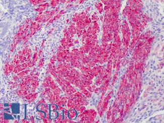
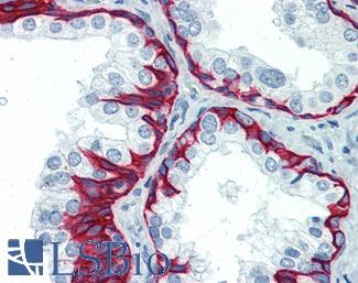
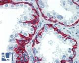
SMMHC
SMMHC (smooth muscle myosin heavy chain / MYH11) is a myoepithelial breast cancer marker. It is overexpressed in HER2-positive, estrogen receptor- and progesterone receptor-negative breast tumors and under-expressed in triple negative breast cancers (Li, 2016). As a myoepithelial marker, MYH11 tends to provide more sensitive results with less myofibroblast cross-reactivity when compared to biomarkers like calponin and smooth muscle actin. It can be used to illustrate ductal wall stability, as ductal tumor cells usually lack expression of MYH11 (Zaha, 2014).
Staining:
Staining for this target is expected to be cytoplasmic.
LSBio's recommended antibody to SMMHC for use in immunohistochemistry is SMMHC Antibody LS‑B5148.
Click here for specifications and further information on this antibody.
Click here for specifications and further information on this antibody.
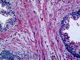
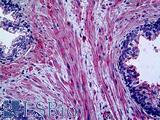 Anti-Myosin, Smooth Muscle Heavy Chain antibody IHC of human prostate. Immunohistochemistry of formalin-fixed, paraffin-embedded tissue after heat-induced antigen retrieval. Antibody LS-B5148 concentration 20 ug/ml.
SMMHC Antibody LS-B5148
CK5 (KRT5)
CK5 (KRT5) is a core basal-like and myoepithelial marker alongside EGFR in the immunohistochemistry of breast cancer (Yang, 2011). It has been found to have increased expression in luminal cells of adenoid cystic carcinoma (Nakai, 2016) and in KRT5 positive, estrogen receptor (ER) and progesterone receptor (PR) negative cells within luminal tumors have been correlated with treatment resistance and increased invasiveness (Axlund, 2013; Kabos, 2011).
Staining:
Staining for this target is expected to be cytoplasmic.
LSBio's recommended antibody to CK5 for use in immunohistochemistry is CK5 Antibody LS‑B3359.
Click here for specifications and further information on this antibody.
Click here for specifications and further information on this antibody.
 Anti-KRT5 / Cytokeratin 5 antibody IHC of human prostate. Immunohistochemistry of formalin-fixed, paraffin-embedded tissue after heat-induced antigen retrieval. Antibody LS-B3359 concentration 10 ug/ml.
CK5 Antibody LS-B3359
CK7 (KRT7)
CK7 (KRT7) is a luminal cell biomarker for breast cancer (Abd El-Rehim, 2004). Cytokeratins are intermediate filaments that are involved in cytoskeletal integrity. This keratin is helpful for distinguishing Paget's disease (breast cancer in the skin) from melanoma. In Paget’s disease, CK7 highlights malignant breast cancer cells in the epidermis (Karakas, 2011; Smith, 1997).
Staining:
Staining for this target is expected to be cytoplasmic.
LSBio's recommended antibody to CK7 for use in immunohistochemistry is CK7 Antibody LS‑B7164.
Click here for specifications and further information on this antibody.
Click here for specifications and further information on this antibody.
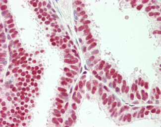
 Anti-KRT7 / Cytokeratin 7 antibody IHC of human placenta. Immunohistochemistry of formalin-fixed, paraffin-embedded tissue after heat-induced antigen retrieval. Antibody LS-B7164 concentration 10 ug/ml.
CK7 Antibody LS-B7164
CK14 (KRT14)
CK14 (KRT14) is a basal-like and myoepithelial marker for normal and cancer tissues of the breast. CK14 expression is linked with metastasis; a study from 2016 by Kevin J. Cheung et al found that metastatic polyclonal tumors shared high levels of KRT14 expression, indicating it as a biomarker for divergent invasive cancers in distant organs that followed an initial “collective invasion” into the stroma (Cheung, 2016). They found a drastic increase in CK14 expression in circulating tumor cells relative to primary tumors. CK14 may thus be an effective target not just as a myoepithelial marker but also for highly invasive metastatic polyclonal breast cancers.
Staining:
Staining for this target is predominantly cytoplasmic.
LSBio's recommended antibody to CK14 for use in immunohistochemistry is CK14 Antibody LS‑B3916.
Click here for specifications and further information on this antibody.
Click here for specifications and further information on this antibody.

 Anti-KRT14 / Cytokeratin 14 antibody IHC of human skin. Immunohistochemistry of formalin-fixed, paraffin-embedded tissue after heat-induced antigen retrieval. Antibody LS-B3916 dilution 1:25.
CK14 Antibody LS-B3916
CK20 (KRT20)
CK20 (KRT20) is an epithelial marker often used together with KRT7 (CK7) when trying to determine a metastatic carcinoma’s point of origin, and a CK7 positive, CK20 negative staining pattern is present in many breast cancers (Tsao, 2007). CK20 may be also be a useful marker for circulating tumor cells in breast cancer patients, and higher levels of expression in the peripheral blood have been found to be correlated with ER-negative high grade tumors and with lymph node metastases (Tunca, 2012; Lasa, 2013). Cytokeratins are intermediate filaments that contribute to cytoskeletal integrity in epithelial cell types, and cytokeratin 20 localizes to the cytoplasm.
Staining:
Staining for this target is expected to be cytoplasmic.
LSBio's recommended antibody to CK20 for use in immunohistochemistry is CK20 Antibody LS‑B10488.
Click here for specifications and further information on this antibody.
Click here for specifications and further information on this antibody.

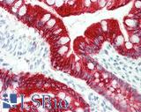 Anti-KRT20 / Cytokeratin 20 antibody IHC staining of human small intestine. Immunohistochemistry of formalin-fixed, paraffin-embedded tissue after heat-induced antigen retrieval. Antibody LS-B10488 dilution 1:200.
CK20 Antibody LS-B10488
E-Cadherin (CDH1)
E-cadherin (CDH1), a cadherin involved in cell to cell adhesion, sees consistent loss of expression in invasive lobular carcinoma of the breast and this coincides with the induction of metastatic and invasive properties in lobular tumors (Ciriello, 2015). CDH1 is often knocked out in ERBB2/HER2- negative, estrogen receptor-positive invasive lobular cancers (Hugo, 2017) and this downregulation is a predictor of poor prognosis (Li, 2017). A study from 2017 looking at the MDA-MB-468 human breast cancer cell line found that e-cadherin knockdown is specifically correlated with invasion, while higher expression profiles are more closely linked with higher proliferation (Ping, 2016), highlighting its utility as a marker in immunohistochemistry staining of invasive lobular tumors.
Staining:
Cadherins typically show membranous staining in immunohistochemistry.
LSBio's recommended antibody to E‑cadherin for use in immunohistochemistry is E‑cadherin Antibody LS‑B4674.
Click here for specifications and further information on this antibody.
Click here for specifications and further information on this antibody.
 Anti-E Cadherin antibody IHC of human liver. Immunohistochemistry of formalin-fixed, paraffin-embedded tissue after heat-induced antigen retrieval. Antibody LS-B4674 concentration 5 ug/ml. This image was taken for the unconjugated form of this product. Other forms have not been tested.
E‑cadherin Antibody LS-B4674
p120 (CTNND1)
p120 (CTNND1) may be used alongside e-cadherin (CDH1) in immunohistochemistry as a marker for lobular breast carcinoma and has been found to be a useful biomarker for early lobular lesions (Dabbs, 2007). Alongside downregulation of e-cadherin, immunohistochemistry staining of p120 shows it to be strongly positive in lobular tumors, with redistribution from the membrane to the cytoplasm (Li, 2014; Li, 2010).
Staining:
Staining for this target is expected to be nuclear.
LSBio's recommended antibody to p120 for use in immunohistochemistry is p120 Antibody LS‑B14422.
Click here for specifications and further information on this antibody.
Click here for specifications and further information on this antibody.
FOXA1
FOXA1 is a transcription factor involved in differentiation that is necessary for estrogen receptor (ER/ESR1) chromatin interaction and regulates many of the same genes. It is a useful marker for luminal breast cancers, as it is often expressed in luminal subtypes independent of ER expression and it does not see expression in basal subtypes (Bernardo, 2013). In ER-negative tumors, a lack of FOXA1 expression is associated with worse prognosis, as loss of FOXA1 is associated with the consequential expression of metastases-prone, basal-specific proteins that it normally acts to directly repress (Bernardo, 2013; Albergaria, 2009; Gong, 2014).
LSBio's recommended antibody to FOXA1 for use in immunohistochemistry is FOXA1 Antibody LS‑B4356.
Click here for specifications and further information on this antibody.
Click here for specifications and further information on this antibody.
 Anti-FOXA1 antibody IHC of human prostate. Immunohistochemistry of formalin-fixed, paraffin-embedded tissue after heat-induced antigen retrieval. Antibody LS-B4356 concentration 5 ug/ml.
FOXA1 Antibody LS-B4356
GATA3
The protein GATA3 is a transcription factor and is a useful biomarker for breast cancer. It has been found to be significantly mutated in luminal breast cancers (Goncalves, 2014), and it is also regularly highly expressed in breast carcinoma. It stains at high levels of expression across many subtypes including triple-negative breast cancers (Kandalaft, 2016) and for both primary and metastatic carcinomas (Sangoi, 2016), and it is considered a biomarker for the Luminal A subtype of epithelial cells (Yersal, 2014).
Staining:
Staining for this target is expected to be nuclear.
LSBio's recommended antibody to GATA3 for use in immunohistochemistry is GATA3 Antibody LS‑B4163.
Click here for specifications and further information on this antibody.
Click here for specifications and further information on this antibody.
 Anti-GATA3 antibody IHC of human breast. Immunohistochemistry of formalin-fixed, paraffin-embedded tissue after heat-induced antigen retrieval. Antibody LS-B4163 dilution 1:200.
GATA3 Antibody LS-B4163
Apoptosis and Angiogenesis Markers In Breast Cancer
VEGFA
VEGFA protein is a marker for tumorigenesis that is involved in angiogenesis and proliferation and is regularly upregulated in breast and other cancers. VEGFA is consistently expressed throughout tumorigenesis and metastasis at higher levels than normal tissue, and seems to be particularly overexpressed in triple-negative breast cancers (Taneja, 2010). VEGFA is a growth factor in vascular endothelial cells, where it activates cellular proliferation, angiogenesis and migration (O’Leary, 2016). VEGFA is involved in activating PI3K-Akt, MEK-ERK and other downstream signaling pathways, and as an actor in angiogenesis it promotes an increase in microvessel density when overexpressed in cancer (Taneja, 2010).
Staining:
Staining for this target is a secreted cytokine, and it can show both cytoplasmic and extracellular staining.
LSBio's recommended antibody to VEGFA for use in immunohistochemistry is VEGFA Antibody LS‑B7747.
Click here for specifications and further information on this antibody.
Click here for specifications and further information on this antibody.
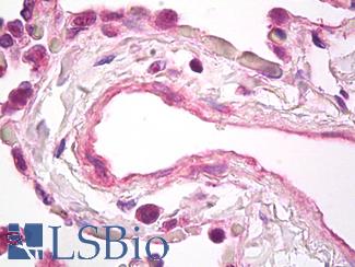
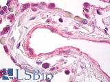 Anti-VEGF antibody IHC of human lung, vascular endothelium. Immunohistochemistry of formalin-fixed, paraffin-embedded tissue after heat-induced antigen retrieval. Antibody LS-B7747 dilution 5 ug/ml.
VEGFA Antibody LS-B7747
BCL2
BCL2 is a marker of apoptosis in breast cancer, where overexpression of this protein is associated with a more favorable prognostic outcome in estrogen receptor- (ER) positive patients. Despite this, the function of BCL2 is anti-apoptotic and it is considered an oncogene (Hwang, 2017), and ER-negative, BCL2-positive patients have been shown to have worse outcomes (Zaha, 2014). It is a useful marker for specifically the luminal A subtype of breast cancer (Yersal, 2014; Eom, 2016). BCL2 staining is typically cytoplasmic, but can also localize to membranes.
LSBio's recommended antibody to BCL2 for use in immunohistochemistry is BCL2 Antibody LS‑B1173.
Click here for specifications and further information on this antibody.
Click here for specifications and further information on this antibody.
 Anti-BCL2 / Bcl-2 antibody IHC of human spleen. Immunohistochemistry of formalin-fixed, paraffin-embedded tissue after heat-induced antigen retrieval. Antibody LS-B1173 concentration 20 ug/ml.
BCL2 Antibody LS-B1173
CD31
CD31 is an angiogenesis marker in breast cancer, and increased staining of vessels within lymph nodes is an indicator of potential lymph node metastases of invasive ductal carcinoma (Popiela, 2008; Zaha, 2014). This protein is expressed in endothelial cells within capillaries and vessels. CD31 is a cellular adhesion molecule, and staining is typically membranous.
LSBio's recommended antibody to CD31 for use in immunohistochemistry is CD31 Antibody LS‑B3446.
Click here for specifications and further information on this antibody.
Click here for specifications and further information on this antibody.
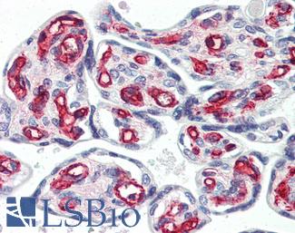
 Anti-CD31 antibody IHC of human placenta. Immunohistochemistry of formalin-fixed, paraffin-embedded tissue after heat-induced antigen retrieval. Antibody LS-B3446 dilution 1:200.
CD31 Antibody LS-B3446
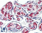
CD34
CD34 is a hematopoietic stem cell marker/adhesion protein that is also expressed in endothelium, where staining is typically membranous. This protein is also occasionally expressed in fibroblasts, and is present in the cytoplasm of mesenchymal cells surrounding benign glandular tissue in the breast. Consistent loss of expression in breast mesenchymal fibroblasts is associated with high grade, malignant breast cancers (Hua, 2011). Loss of expression of this protein in the stroma coincides with expression of smooth muscle actin and the adoption of invasive properties in ductal tumor cells (Catteau, 2013).
LSBio's recommended antibody to CD34 for use in immunohistochemistry is CD34 Antibody LS‑B2652.
Click here for specifications and further information on this antibody.
Click here for specifications and further information on this antibody.
 Anti-CD34 antibody IHC of human placenta. Immunohistochemistry of formalin-fixed, paraffin-embedded tissue after heat-induced antigen retrieval. Antibody LS-B2652 concentration 10 ug/ml.
CD34 Antibody LS-B2652
Metastatic Markers In Breast Cancer
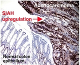
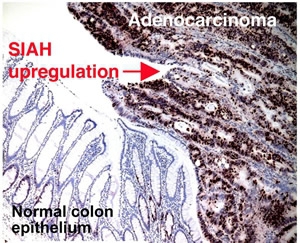
SIAH2
SIAH2 is a novel target in breast and other cancers; in a study on advanced and metastatic breast cancer, Siewertz van Reesama et al found that both SIAH and EGFR outperform standard breast cancer biomarkers in predicting advanced stages of metastasis. SIAH2 is a downstream member of the RAS pathway, where it is required for signal transduction (Van Reesema, 2016). SIAH2 expression acts as a sign of activation of the RAS pathway (Van Reesema, 2016), and its deficiency has been found to reduce MAPK signaling (Ahmed, 2008). SIAH2 has been found to be overexpressed in invasive breast cancers versus ductal carcinomas and normal tissue, where it may cause aberrant activation of the ERK pathway, encouraging proliferation and simultaneous dysregulation of apoptotic signaling (Sun, 2016).
Staining:
Staining for this target can be both cytoplasmic and nuclear.
LSBio's recommended antibody to SIAH2 for use in immunohistochemistry is SIAH2 Antibody LS‑C141889.
Click here for specifications and further information on this antibody.
Click here for specifications and further information on this antibody.
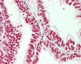
Prolactin-induced protein (PIP / GCDFP-15)
Prolactin-induced protein (PIP / GCDFP-15) is a secreted protein produced by benign and malignant salivary gland and breast tissue. PIP is overexpressed in many breast carcinomas, where increased expression has been found to increase the cell adhesive and proliferative properties of malignant cells (Vanneste 2015; Naderi 2015).
Staining:
This target typically stains secretions (may appear cytoplasmic, membranous, and/or extracellular).
LSBio's recommended antibody to PIP / GCDFP‑15 for use in immunohistochemistry is PIP / GCDFP‑15 Antibody LS‑B2498.
Click here for specifications and further information on this antibody.
Click here for specifications and further information on this antibody.

 Anti-PIP / GCDFP15 antibody IHC of human breast. Immunohistochemistry of formalin-fixed, paraffin-embedded tissue after heat-induced antigen retrieval. Antibody LS-B2498 concentration 5 ug/ml.
PIP / GCDFP‑15 Antibody LS-B2498
Mammaglobin A (SCGB2A2)
Mammaglobin A (SCGB2A2) is a breast cancer biomarker that is tissue specific to breast and highly expressed in breast cancers. Overexpression of this protein has been correlated with proliferative and invasive properties in tumor cells (Picot, 2016), and coincides with simultaneous upregulation of Ki-67 (MKI67) protein – these two markers combined can be a useful pair in tissue specific immunohistochemistry staining of breast tumors (Gargano, 2006).
Staining:
Staining for this target is has been demonstrated to be cytoplasmic and in secretions.
LSBio's recommended antibody to SCGB2A2 for use in immunohistochemistry is SCGB2A2 Antibody LS‑B3007.
Click here for specifications and further information on this antibody.
Click here for specifications and further information on this antibody.
 Anti-SCGB2A2 / Mammaglobin A antibody IHC of human breast. Immunohistochemistry of formalin-fixed, paraffin-embedded tissue after heat-induced antigen retrieval. Antibody LS-B3007 concentration 4 ug/ml.
SCGB2A2 Antibody LS-B3007
TNBC Markers In Breast Cancer
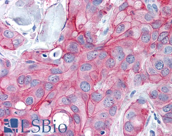
PIM1
PIM1 is a marker in breast tumors with particular relevance as a critical proliferative regulator in triple-negative breast cancers (TNBC). PIM1 was found to regulate proliferation and apoptosis in TNBC, with overexpression that correlates with MYC-target transcription and tumor proliferation. It is a potential novel drug target in TNBC treatment (Brasó-Maristany, 2016).
Staining:
Staining for PIM1 is expected to be cytoplasmic.
LSBio's recommended antibody to PIM1 for use in immunohistochemistry is PIM1 Antibody LS‑B5493.
Click here for specifications and further information on this antibody.
Click here for specifications and further information on this antibody.
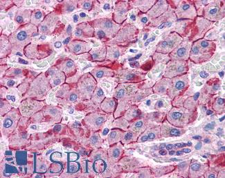
 Anti-PIM1 antibody IHC of human adrenal. Immunohistochemistry of formalin-fixed, paraffin-embedded tissue after heat-induced antigen retrieval. Antibody LS-B5493 concentration 5 ug/ml.
PIM1 Antibody LS-B5493
References ▷
-
Abd El-Rehim, Dalia M et al. Expression of luminal and basal cytokeratins in human breast carcinoma The Journal of Pathology 2004 Jun; 203.2; 661-671
[Full Text Article] [Pubmed: 15141381] -
Ahmed, Atique U et al. Effect of Disrupting Seven-in-Absentia Homolog 2 Function on Lung Cancer Cell Growth JNCI Journal of the National Cancer Institute 2008 Nov; 100.22; 1606-1629
[Full Text Article] [Pubmed: 19001609] [PMC: PMC2720765] -
Albergaria, André et al. Expression of FOXA1 and GATA-3 in breast cancer: the prognostic significance in hormone receptor-negative tumours Breast Cancer Research 2009 Jun; 11; R40
[Full Text Article] -
Alexandrov, Ludmil B et al. Clock-like mutational processes in human somatic cells Nature genetics 2015 Dec; 47.12; 1402-1407
[Full Text Article] [Pubmed: 26551669] [PMC: PMC4783858] -
Anantha, Rachel W et al. Functional and mutational landscapes of BRCA1 for homology-directed repair and therapy resistance eLife 2017 ; 6; e21350
[Full Text Article] [Pubmed: 28398198] [PMC: PMC5432210] -
Axlund, Sunshine Daddario et al. Progesterone-inducible cytokeratin 5 positive cells in luminal breast cancer exhibit progenitor properties Hormones & cancer 2013 Feb; 4.1; 36-49
[Full Text Article] [Pubmed: 23184698] [PMC: PMC3549640] -
Balmaña, J et al. BRCA in breast cancer: ESMO Clinical Recommendations Annals of Oncology 2009 May; 20.suppl_4; iv19-iv20
[Full Text Article] -
Bernardo GM1 et al. FOXA1 represses the molecular phenotype of basal breast cancer cells Oncogene 2013 Jan; 32.5; 554-63
[Full Text Article] [Pubmed: 22391567] [PMC: PMC3371315] -
Brasó-Maristany, Fara et al. PIM1 kinase regulates cell death, tumor growth and chemotherapy response in triple-negative breast cancer Nature medicine 2016 Nov; 22.11; 1303-1313
[Full Text Article] [Pubmed: 27775704] [PMC: PMC5552044] -
Burns, Michael B et al. Evidence for APOBEC3B mutagenesis in multiple human cancers Nature genetics 2013 Sep; 45.9; 977-983
[Full Text Article] [Pubmed: 23852168] [PMC: PMC3902892] -
Campbell, Joshua D et al. Distinct patterns of somatic genome alterations in lung adenocarcinomas and squamous cell carcinomas Nature Genetics 2016 Jun; 48.6; 607
[Full Text Article] -
Catteau, Xavier et al. Variable Stromal Periductular Expression of CD34 and Smooth Muscle Actin (SMA) in Intraductal Carcinoma of the Breast PLOS ONE 2013 Mar; 8.3; e57773
[Full Text Article] -
Changavi, Arathi A et al. Epidermal Growth Factor Receptor Expression in Triple Negative and Nontriple Negative Breast Carcinomas Journal of Laboratory Physicians 2015 Jul; 7.2; 79-83
[Full Text Article] [Pubmed: 26417156] [PMC: PMC4559633] -
Cheung, Kevin J et al. Polyclonal breast cancer metastases arise from collective dissemination of keratin 14-expressing tumor cell clusters Proceedings of the National Academy of Sciences of the United States of America 2016 Feb; 113.7; E854-E863
[Full Text Article] [Pubmed: 26831077] [PMC: PMC4763783] -
Ciriello, Giovanni et al. Comprehensive Molecular Portraits of Invasive Lobular Breast Cancer Cell 2015 Jan; 163.2; 506-519
[Full Text Article] [Pubmed: 26451490] -
Dabbs, David J et al. Lobular versus ductal breast neoplasms: the diagnostic utility of p120 catenin The American Journal of Surgical Pathology 2007 Mar; 31.3; 427-437
[Full Text Article] [Pubmed: 17325485] -
Decker, Brennan et al. Biallelic BRCA2 Mutations Shape the Somatic Mutational Landscape of Aggressive Prostate Tumors American Journal of Human Genetics 2016 May; 98.5; 818-829
[Full Text Article] [Pubmed: 27087322] [PMC: PMC4863563] -
Eom, Yong Hwa et al. BCL2 as a Subtype-Specific Prognostic Marker for Breast Cancer Journal of Breast Cancer 2016 Sep; 19.3; 252-260
[Full Text Article] [Pubmed: 27721874] [PMC: PMC5053309] -
Foo, T K et al. Compromised BRCA1-PALB2 interaction is associated with breast cancer risk Oncogene 2017 Jul; 36.29; 4161-4170
[Full Text Article] [Pubmed: 28319063] [PMC: PMC5519427] -
Gargano, G et al. Detection and quantification of mammaglobin in the blood of breast cancer patients: can it be useful as a potential clinical marker? Preliminary results of a GOIM (Gruppo Oncologico dell'Italia Meridionale) prospective study Annals of Oncology: Official Journal of the European Society for Medical Oncology 2006 Jun; 17 Suppl 7; vii41-45
[Full Text Article] [Pubmed: 16760290] -
Gaynor, Katherine U et al. GATA3 Mutations Found in Breast Cancers May Be Associated with Aberrant Nuclear Localization, Reduced Transactivation and Cell Invasiveness Hormones and Cancer 2013 Jun; 4.3; 123-139
[Full Text Article] -
Godet, Inês et al. BRCA1 and BRCA2 mutations and treatment strategies for breast cancer Integrative cancer science and therapeutics 2017 Feb; 4.1;
[Pubmed: 28706734] [PMC: PMC5505673] -
Goncalves, Rodrigo et al. New concepts in breast cancer genomics and genetics Breast cancer research: BCR 2014 ; 16.5; 460
[Pubmed: 25606588] [PMC: PMC4384360] -
Gong, C et al. FOXA1 repression is associated with loss of BRCA1 and increased promoter methylation and chromatin silencing in breast cancer Oncogene 2015 Sep; 34.39; 5012
[Full Text Article] -
Hause, Ronald J et al. Classification and characterization of microsatellite instability across 18 cancer types Nature Medicine 2016 Nov; 22.11; 1342
[Full Text Article] -
Hua, Xing et al. Expression and role of fibroblast activation protein-alpha in microinvasive breast carcinoma Diagnostic Pathology 2011 Nov; 6; 111
[Full Text Article] -
Hugo, H J et al. Epithelial requirement for in vitro proliferation and xenograft growth and metastasis of MDA-MB-468 human breast cancer cells: oncogenic rather than tumor-suppressive role of E-cadherin Breast Cancer Research : BCR 2017 ; 19;
[Full Text Article] [Pubmed: 28750639] [PMC: PMC5530912] -
Hwang, Joyce K et al. Related Mechanisms of Antibody Somatic Hypermutation and Class Switch Recombination Microbiology spectrum 2015 Feb; 3.1;
[Full Text Article] [Pubmed: 26104555] [PMC: PMC4481323] -
Hwang, Ki-Tae et al. Prognostic Influence of BCL2 on Molecular Subtypes of Breast Cancer Journal of Breast Cancer 2017 Mar; 20.1; 54-64
[Full Text Article] [Pubmed: 28382095] [PMC: PMC5378580] -
Inic, Zorka et al. Difference between Luminal A and Luminal B Subtypes According to Ki-67, Tumor Size, and Progesterone Receptor Negativity Providing Prognostic Information Clinical Medicine Insights. Oncology 2014 Sep; 8; 107-111
[Full Text Article] [Pubmed: 25249766] [PMC: PMC4167319] -
Jeon, Myeongjin et al. Dimerization of EGFR and HER2 induces breast cancer cell motility through STAT1-dependent ACTA2 induction Oncotarget 2016 Jul; 8.31; 50570-50581
[Full Text Article] [Pubmed: 28881584] [PMC: PMC5584169] -
Jeselsohn, Rinath et al. ESR1 mutations as a mechanism for acquired endocrine resistance in breast cancer Nature reviews. Clinical oncology 2015 Jan; 12.1; 573-583
[Full Text Article] [Pubmed: 26122181] [PMC: PMC4911210] -
Kabos, Peter et al. Cytokeratin 5 positive cells represent a steroid receptor negative and therapy resistant subpopulation in luminal breast cancers Breast Cancer Research and Treatment 2011 Jul; 128.1; 45-55
[Full Text Article] [Pubmed: 20665103] [PMC: PMC3851293] -
Kandalaft, Patricia L et al. Comparative Sensitivities and Specificities of Antibodies to Breast Markers GCDFP-15, Mammaglobin A, and Different Clones of Antibodies to GATA-3: A Study of 338 Tumors Using Whole Sections Applied immunohistochemistry & molecular morphology: AIMM 2016 Jan; 24.9; 609-614
[Full Text Article] [Pubmed: 26447897] -
Kanu, Nnennaya et al. DNA replication stress mediates APOBEC3 family mutagenesis in breast cancer Genome Biology 2016 Sep; 17;
[Full Text Article] [Pubmed: 27634334] [PMC: PMC5025597] -
Karakas, Cansu et al. Paget's disease of the breast Journal of Carcinogenesis 2011 Dec; 10;
[Full Text Article] [Pubmed: 22279416] [PMC: PMC3263015] -
Lada, Artem G et al. AID/APOBEC cytosine deaminase induces genome-wide kataegis Biology Direct 2012 Dec; 7; 47
[Full Text Article] [Pubmed: 23249472] [PMC: PMC3542020] -
Lasa, Adriana et al. Molecular Detection of Peripheral Blood Breast Cancer mRNA Transcripts as a Surrogate Biomarker for Circulating Tumor Cells PLoS ONE 2013 Sep; 8.9;
[Full Text Article] [Pubmed: 24058517] [PMC: PMC3776801] -
Li, Lan et al. [Expression pattern of E-cadherin and p120-catenin in infiltrating lobular carcinoma and ductal carcinoma of the breast and its significance] Zhonghua Zhong Liu Za Zhi [Chinese Journal of Oncology] 2010 Apr; 32.4; 273-277
[Pubmed: 20510078] -
Li, Xiaoxian et al. Diagnostic utility of E-cadherin and P120 catenin cocktail immunostain in distinguishing DCIS from LCIS International Journal of Clinical and Experimental Pathology 2014 Apr; 7.5; 2551-2557
[Pubmed: 24966968] [PMC: PMC4069889] -
Li, Yang et al. Exploring the intrinsic differences among breast tumor subtypes defined using immunohistochemistry markers based on the decision tree Scientific Reports 2016 Jan; 6; 35773
[Full Text Article] -
Li, Zhan et al. Prognostic value of reduced E-cadherin expression in breast cancer: a meta-analysis Oncotarget 2017 Jan; 8.1; 16445-16455
[Full Text Article] [Pubmed: 28147315] [PMC: PMC5369975] -
Liu, Haiyan et al. Application of immunohistochemistry in breast pathology: a review and update Archives of Pathology & Laboratory Medicine 2014 Dec; 138.12; 1629-1642
[Full Text Article] [Pubmed: 25427042] -
Masuda H et al. Role of epidermal growth factor receptor in breast cancer Breast Cancer Research and Treatment 2012 Nov; 136.2; 331-345
[Full Text Article] [Pubmed: 23073759] [PMC: PMC3832208] -
McBride, Kevin M et al. Regulation of class switch recombination and somatic mutation by AID phosphorylation The Journal of Experimental Medicine 2008 Jan; 205.11; 2585-2594
[Full Text Article] [Pubmed: 18838546] [PMC: PMC2571933] -
Middlebrooks, Candace D et al. Association of germline variants in the APOBEC3region with cancer risk and enrichment with APOBEC-signature mutations in tumors Nature Genetics 2016 Nov; 48.11; 1330
[Full Text Article] -
Mohammed, Hisham et al. Progesterone receptor modulates estrogen receptor-α action in breast cancer Nature 2015 Jul; 523.756; 313-317
[Full Text Article] [Pubmed: 26153859] [PMC: PMC4650274] -
Morganella, Sandro et al. The topography of mutational processes in breast cancer genomes Nature Communications 2016 May; 7; 11383
[Full Text Article] -
Naderi, Ali et al. Prolactin-induced protein in breast cancer Advances in Experimental Medicine and Biology 2015 ; 846; 189-200
[Full Text Article] [Pubmed: 25472539] - Nakai, Tokiko et al. The unique luminal staining pattern of cytokeratin 5/6 in adenoid cystic carcinoma of the breast may aid in differentiating it from its mimickers Virchows Archiv 2016 Aug; 469.2; 213-222
-
Nekulova, Marta et al. ΔNp63α expression induces loss of cell adhesion in triple-negative breast cancer cells BMC Cancer 2016 Jan; 16;
[Full Text Article] [Pubmed: 27724925] [PMC: PMC5057421] -
Nik-Zainal, Serena et al. Mutational processes molding the genomes of 21 breast cancers Cell 2012 May; 149.5; 979-993
[Full Text Article] [Pubmed: 22608084] [PMC: PMC3414841] -
Nik-Zainal, Serena et al. Landscape of somatic mutations in 560 breast cancer whole-genome sequences Nature 2016 ; 534.7605; 47-54
[Full Text Article] [Pubmed: 27135926] [PMC: PMC4910866] -
Nik-Zainal, Serena et al. Mutational Signatures in Breast Cancer: The Problem at the DNA Level Clinical cancer research : an official journal of the American Association for Cancer Research 2017 Jun; 23.11; 2617-2629
[Full Text Article] [Pubmed: 28572256] [PMC: PMC5458139] -
O'Kelly, James et al. Functional domains of CCN1 (Cyr61) regulate breast cancer progression International Journal of Oncology 2008 Jul; 33.1; 59-67
[Pubmed: 18575751] -
O'Leary, Nuala A et al. Reference sequence (RefSeq) database at NCBI: current status, taxonomic expansion, and functional annotation Nucleic Acids Research 2016 Jan; 44.D1; D733-745
[Full Text Article] [Pubmed: 26553804] [PMC: PMC4702849] -
Pan, Yunbao et al. P53 and Ki-67 as prognostic markers in triple-negative breast cancer patients PLOS ONE 2017 Feb; 12.2; e0172324
[Full Text Article] -
Periyasamy, Manikandan et al. APOBEC3B-Mediated Cytidine Deamination Is Required for Estrogen Receptor Action in Breast Cancer Cell Reports 2015 Jan; 13.1; 108-121
[Full Text Article] [Pubmed: 26411678] -
Picot, Nadia et al. Mammaglobin 1 promotes breast cancer malignancy and confers sensitivity to anticancer drugs Molecular Carcinogenesis 2016 ; 55.7; 1150-1162
[Full Text Article] [Pubmed: 26207726] -
Ping, Zheng et al. ERBB2 mutation is associated with a worse prognosis in patients with CDH1 altered invasive lobular cancer of the breast Oncotarget 2016 Nov; 7.49; 80655-80663
[Full Text Article] [Pubmed: 27811364] [PMC: PMC5340256] -
Polak, Paz et al. A mutational signature reveals alterations underlying deficient homologous recombination repair in breast cancer Nature Genetics 2017 Jan; 49.1; 1476
[Full Text Article] -
Popiela, Tadeusz J et al. The analysis of CD34 antigen immunoreactivity level in invasive ductal breast cancer with respect to the presence of lymph node metastases Neuro Endocrinology Letters 2008 Aug; 29.4; 443-446
[Pubmed: 18766141] -
Ripperger, Tim et al. Breast cancer susceptibility: current knowledge and implications for genetic counselling European Journal of Human Genetics 2009 Jun; 17.6; 722-731
[Full Text Article] [Pubmed: 19092773] [PMC: PMC2947107] -
Roberts, Maegan E et al. MSH6 and PMS2 germ-line pathogenic variants implicated in Lynch syndrome are associated with breast cancer Genetics in Medicine 2018 Jan; ;
[Full Text Article] -
Romagnolo, Alberto P G et al. BRCA1 as target for breast cancer prevention and therapy Anti-Cancer Agents in Medicinal Chemistry 2015 ; 15.1; 43204
[Pubmed: 25329591] -
Sangoi, Ankur R et al. The Novel Marker GATA3 is Significantly More Sensitive Than Traditional Markers Mammaglobin and GCDFP15 for Identifying Breast Cancer in Surgical and Cytology Specimens of Metastatic and Matched Primary Tumors Applied immunohistochemistry & molecular morphology: AIMM 2016 Apr; 24.4; 229-237
[Full Text Article] [Pubmed: 25906123] [PMC: PMC5610542] -
Smith, K J et al. Cytokeratin 7 staining in mammary and extramammary Paget's disease. Modern pathology : an official journal of the United States and Canadian Academy of Pathology, Inc 1997 Nov; 10.11; 1069-1074
[Pubmed: 9388055] - Sun, Jie et al. Overexpression of seven in absentia homolog 2 protein in human breast cancer tissues is associated with the promotion of tumor cell malignant behavior in in vitro Oncology Reports 2016 Sep; 36.3; 1301-1312
-
Taneja, Pankaj et al. Classical and Novel Prognostic Markers for Breast Cancer and their Clinical Significance Clinical Medicine Insights. Oncology 2010 Apr; 4; 15-34
[Pubmed: 20567632] [PMC: PMC2883240] -
Tanino, Hirokazu et al. BRCAness and Prognosis in Triple-Negative Breast Cancer Patients Treated with Neoadjuvant Chemotherapy PLOS ONE 2016 Dec; 11.12; e0165721
[Full Text Article] -
Tsao, Shu-Chuan et al. Use of caveolin-1, thyroid transcription factor-1, and cytokeratins 7 and 20 in discriminating between primary and secondary pulmonary adenocarcinoma from breast or colonic origin The Kaohsiung Journal of Medical Sciences 2007 Jul; 23.7; 325-331
[Full Text Article] [Pubmed: 17606426] -
Tunca, Berrin et al. CK19, CK20, EGFR and HER2 status of circulating tumor cells in patients with breast cancer Tumori 2012 Apr; 98.2; 243-251
[Full Text Article] [Pubmed: 22677992] -
van Reesema, Lauren L Siewertsz et al. SIAH and EGFR, Two RAS Pathway Biomarkers, are Highly Prognostic in Locally Advanced and Metastatic Breast Cancer EBioMedicine 2016 Aug; 11; 183-198
[Full Text Article] [Pubmed: 27569656] [PMC: PMC5049993] -
Vanneste, Marion et al. Prolactin-Induced Protein regulates cell adhesion in breast cancer Biochemical and Biophysical Research Communications 2015 Dec; 468.4; 850-856
[Full Text Article] [Pubmed: 26585492] -
Verma, I M et al. Rel/NF-kappa B/I kappa B family: intimate tales of association and dissociation Genes & Development 1995 Nov; 9.22; 2723-2735
[Pubmed: 7590248] -
Volinia, Stefano et al. The ubiquitous 'cancer mutational signature' 5 occurs specifically in cancers with deleted FHIT alleles Oncotarget 2017 Nov; 8.6; 102199-102211
[Full Text Article] [Pubmed: 29254236] [PMC: PMC5731946] -
Vos, Shoko et al. BRCA promoter methylation in sporadic versus BRCA germline mutation-related breast cancers Breast Cancer Research : BCR 2017 ; 19;
[Full Text Article] [Pubmed: 28569220] [PMC: PMC5452400] -
Yang, Xiaohong R et al. Associations of Breast Cancer Risk Factors With Tumor Subtypes: A Pooled Analysis From the Breast Cancer Association Consortium Studies JNCI Journal of the National Cancer Institute 2011 Feb; 103.3; 250-263
[Full Text Article] [Pubmed: 21191117] [PMC: PMC3107570] -
Yersal, Ozlem et al. Biological subtypes of breast cancer: Prognostic and therapeutic implications World Journal of Clinical Oncology 2014 Aug; 5.3; 412-424
[Full Text Article] [Pubmed: 25114856] [PMC: PMC4127612] -
Zaha, Dana Carmen et al. Significance of immunohistochemistry in breast cancer World Journal of Clinical Oncology 2014 Aug; 5.3; 382-392
[Full Text Article] [Pubmed: 25114853] [PMC: PMC4127609] -
Zhao, Wen et al. PIM1: a promising target in patients with triple-negative breast cancer Medical Oncology 2017 Aug; 34.8; 142
[Full Text Article]


