Login
Registration enables users to use special features of this website, such as past
order histories, retained contact details for faster checkout, review submissions, and special promotions.
order histories, retained contact details for faster checkout, review submissions, and special promotions.
Forgot password?
Registration enables users to use special features of this website, such as past
order histories, retained contact details for faster checkout, review submissions, and special promotions.
order histories, retained contact details for faster checkout, review submissions, and special promotions.
Quick Order
Products
Antibodies
ELISA and Assay Kits
Research Areas
Infectious Disease
Resources
Purchasing
Reference Material
Contact Us
Location
Corporate Headquarters
Vector Laboratories, Inc.
6737 Mowry Ave
Newark, CA 94560
United States
Telephone Numbers
Customer Service: (800) 227-6666 / (650) 697-3600
Contact Us
Additional Contact Details
Login
Registration enables users to use special features of this website, such as past
order histories, retained contact details for faster checkout, review submissions, and special promotions.
order histories, retained contact details for faster checkout, review submissions, and special promotions.
Forgot password?
Registration enables users to use special features of this website, such as past
order histories, retained contact details for faster checkout, review submissions, and special promotions.
order histories, retained contact details for faster checkout, review submissions, and special promotions.
Quick Order
| Catalog Number | Size | Price |
|---|---|---|
| LS-C204558-100 | 100 µl | $376 |
1 of 2
2 of 2
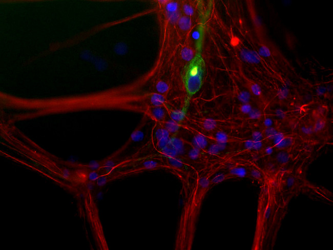
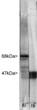
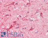
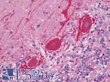
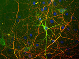
Polyclonal Rabbit anti‑Human NF‑L / NEFL Antibody (IHC, IF, WB) LS‑C204558
Polyclonal Rabbit anti‑Human NF‑L / NEFL Antibody (IHC, IF, WB) LS‑C204558
Antibody:
NF-L / NEFL Rabbit anti-Human Polyclonal Antibody
Application:
IHC, ICC, IF, WB
Reactivity:
Human, Rat
Format:
Unconjugated, Unmodified
Toll Free North America
 (800) 227-6666
For Research Use Only
Overview
Antibody:
NF-L / NEFL Rabbit anti-Human Polyclonal Antibody
Application:
IHC, ICC, IF, WB
Reactivity:
Human, Rat
Format:
Unconjugated, Unmodified
Specifications
Description
NEFL antibody LS-C204558 is an unconjugated rabbit polyclonal antibody to NEFL (NF-L) from human. It is reactive with human and rat. Validated for ICC, IF, IHC and WB.
Target
Human NF-L / NEFL
Synonyms
NEFL | 68 kDa neurofilament protein | CMT1F | CMT2E | NF68 | NF-L | NFL | Neurofilament subunit NF-L | PPP1R110 | Neurofilament light polypeptide | CMTDIG
Host
Rabbit
Reactivity
Human, Rat
(tested or 100% immunogen sequence identity)
Clonality
Polyclonal
Conjugations
Unconjugated
Purification
Antiserum
Modifications
Unmodified
Immunogen
Pig intermediate filaments were prepared from spinal cords by the method of Delacourte et al., and this cytoskeletal material was dissolved in 6M urea. NF-L was purified by ion exchange chromatography, followed by preparative gel electrophoresis.
Specificity
Human NF-L / NEFL
Applications
- IHC
- ICC (1:5000)
- Immunofluorescence (1:500 - 1:1000)
- Western blot (1:20000)

|
Performing IHC? See our complete line of Immunohistochemistry Reagents including antigen retrieval solutions, blocking agents
ABC Detection Kits and polymers, biotinylated secondary antibodies, substrates and more.
|
Usage
Try at dilutions of 1:500-1:1000 for immunofluorescence, and 1:5000 for ABC or other enzyme linked immunocytochemical procedures. For western blots try at 1:20000.
Presentation
Antiserum
Storage
Store at 4°C or -20°C. Avoid freeze-thaw cycles.
Restrictions
For research use only. Intended for use by laboratory professionals.
About NF-L / NEFL
Publications (0)
Customer Reviews (0)
Featured Products
Species:
Human
Applications:
IHC, IHC - Paraffin, IHC - Frozen, Western blot, Immunoprecipitation, ELISA
Species:
Human
Applications:
IHC, IHC - Paraffin, Western blot
Species:
Human
Applications:
IHC, IHC - Paraffin, ICC, Immunofluorescence, Western blot
Species:
Human, Mouse, Rat
Applications:
IHC, IHC - Paraffin, Peptide Enzyme-Linked Immunosorbent Assay
Species:
Human, Mouse, Rat, Bovine, Pig
Applications:
ICC, Immunofluorescence, Western blot
Request SDS/MSDS
To request an SDS/MSDS form for this product, please contact our Technical Support department at:
Technical.Support@LSBio.com
Requested From: United States
Date Requested: 11/22/2024
Date Requested: 11/22/2024


