Products
Research Areas
COVID-19
Resources
Login
Quick Order
Cart Cart lightblue
Login
Registration enables users to use special features of this website, such as past
order histories, retained contact details for faster checkout, review submissions, and special promotions.


Fields marked with a * are required.

Login
Quick Order
Contact Us

Location


Corporate Headquarters

Vector Laboratories, Inc.
6737 Mowry Ave
Newark, CA 94560
United States

Telephone Numbers


Customer Service: (800) 227-6666 / (650) 697-3600


Contact Us


Additional Contact Details

Login
Registration enables users to use special features of this website, such as past
order histories, retained contact details for faster checkout, review submissions, and special promotions.


Fields marked with a * are required.

Login
Quick Order

GDF15

growth differentiation factor 15

Growth differentiation factor 15 (GDF15) is a protein belonging to the transforming growth factor beta superfamily that has a role in regulating inflammatory and apoptotic pathways in injured tissues and during disease processes. GDF15 is also known as TGF-PL, MIC-1, PDF, PLAB, and PTGFB. GDF15 mRNA is most abundant in the liver, with lower levels seen in some other tissues. Its expression in liver can be significantly up-regulated in during injury of organs such as liver, kidney, heart and lung.

Gene Name: growth differentiation factor 15
Family/Subfamily: TGF beta , not assigned-TGF beta
Synonyms: GDF15, MIC1, MIC-1, NAG-1, NRG-1, NSAID-regulated gene 1 protein, PLAB, NSAID-activated gene 1 protein, Placental TGF-beta, PTGFB, GDF-15, PTGF-beta
Target Sequences: NM_004864 NP_004855.2 Q99988

Publications (1)
1
The myokine GDF-15 is a potential biomarker for myositis and associates with the protein aggregates of sporadic inclusion body myositis. Boel De Paepe, Fien Verhamme, Jan L De Bleecker. Cytokine. 2020 March;127:154966. [Full Text Article] [PubMed:31901761] Related Antibodies: LS-B2413.

☰ Filters
Antibodies (100)
ELISA Kits (36)
Proteins (11)
Competitive EIA (4)
Custom (2)
Development Kit (1)
Over-Expression Lysate (2)
Primary (100)
Recombinant (9)
Sandwich (23)
Sandwich CLIA (6)
GDF15 (147)
Human (95)
Mouse (36)
Rat (29)
Chicken (1)
Guinea pig (1)
Monkey (2)
Pig (1)
Porcine (1)
IHC (29)
IHC-P (6)
WB (93)
Flo (8)
ELISA (49)
IF (1)
Peptide-ELISA (4)
rabbit (86)
mouse (11)
goat (1)
rat (2)
Product Group
DevKit DuoPlus (1)
IHCPlus (3)
PathPlus Other (2)
IgG (49)
IgG1 (2)
IgG1,k (4)
IgG2a,l (2)
IgG2b,k (2)
monoclonal mc (13)
polyclonal pc (87)
09 (1)
12 (1)
23B3.D2.H5 (2)
23G10.E4.F8 (2)
44 (1)
5E1A5 (1)
6D12.H10.E4 (2)
7C12.B3.F2 (2)
ME-6D10 (1)
12 x 8-Well Microstrips (3)
96-Well Microplate (6)
96-Well Strip Plate (25)
Antibody Pair and Standard (1)
APC (4)
APC Conjugated (5)
Biotin Conjugated (12)
Cy3 Conjugated (4)
Cy7 Conjugated (4)
FITC Conjugated (7)
HRP Conjugated (13)
PE Conjugated (5)
Preservative Free (3)
Unconjugated (50)
aa189-303 (8)
aa193-296 (8)
aa195-308 (8)
aa30-308 (8)
aa51-79 (7)
C-Terminus (5)
aa35-84 (4)
1-80 aa, Internal (1)
Internal (1)
aa15-64 (1)
aa258-293 (1)
aa286-299 (1)
aa31-80 (1)
No (146)
Yes (1)
Cell Culture Supernatants (9)
Cell Lysates (4)
EDTA Plasma (1)
Heparin Plasma (1)
Plasma (29)
Serum (31)
Tissue Homogenates (7)
GST (1)
His (4)
His, N-Terminal (2)
His-S (1)
Myc-DDK (Flag) (2)
Human (7)
Mouse (2)
Rat (1)
293T Cells (1)
CHO Cells (1)
E. coli (7)
HEK 293 Cells (1)
Wheat Germ Extract (1)
Purified (2)
Low endotoxin level (2)
Biologically active (1)
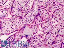
GDF15 Antibody - Anti-GDF15 antibody IHC of human placenta. Immunohistochemistry of formalin-fixed, paraffin-embedded tissue after heat-induced antigen retrieval. Antibody concentration 5 ug/ml.
Select
GDF15 Goat anti-Human Polyclonal (aa286-299) Antibody
Human
Flo, IHC, IHC-P, Peptide-ELISA, WB
Unconjugated
50 µg/$485
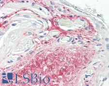
GDF15 Antibody - Anti-GDF15 antibody IHC of human placenta. Immunohistochemistry of formalin-fixed, paraffin-embedded tissue after heat-induced antigen retrieval. Antibody dilution 10 ug/ml.
Select
GDF15 Rat anti-Human Monoclonal (C-Terminus) (6D12.H10.E4) Antibody
Mouse, Human
ELISA, IHC, IHC-P, WB
Unconjugated
50 µg/$460
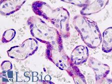
GDF15 Antibody - Anti-GDF15 antibody IHC staining of human placenta. Immunohistochemistry of formalin-fixed, paraffin-embedded tissue after heat-induced antigen retrieval.
Select
GDF15 Rabbit anti-Human Polyclonal (aa51-79) Antibody
Human
Flo, IHC, IHC-P, WB
Unconjugated
200 µl/$460
Select
GDF15 Mouse anti-Human Monoclonal (ME-6D10) Antibody
Human
ELISA, Flo
Unconjugated
50 µg/$536; 100 µg/$836
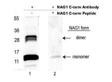
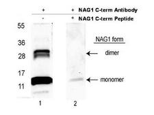
GDF15 Antibody - Anti-NAG-1/GDF15 (C-terminal) Antibody - Western Blot. Western blot of affinity purified anti-NAG-1/GDF15 (C-terminal) antibody shows detection NAG-1 purified from CHO cells as a 14 kD band corresponding to monomer and a 28 kD band corresponding to dimerized NAG-1. Samples were electrophoresed on a 4-20% gradient gel under reducing conditions. Lane 1 shows NAG-1 detection. Lane 2 shows reactivity is blocked when this antibody is pre-incubated with the immunizing peptide prior to Western blotting. This image was taken for the unconjugated form of this product. Other forms have not been tested.
Select
GDF15 Rabbit anti-Human Polyclonal (Biotin) Antibody
Mouse, Human
ELISA, IHC, WB
Biotin Conjugated
100 µg/$544
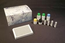
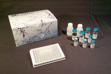
GDF15 ELISA Kit
Select
Sandwich
96-Well Microplate
Mouse
15.6 - 1000 pg/ml
Colorimetric - 450nm (TMB)
Cell Culture Supernatants, Cell Lysates, Plasma, Serum
1 Plate/$699
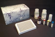
GDF15 ELISA Kit
Select
Sandwich
12 x 8-Well Microstrips
Human
8 - 500 pg/ml
Colorimetric - 450nm (TMB)
1 Plate/$534
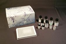
GDF15 ELISA Kit
Select
Sandwich
96-Well Strip Plate
Mouse
0.156 - 100 ng/ml
Colorimetric - 450nm (TMB)
Plasma, Serum, Tissue Homogenates
1 Plate/$748
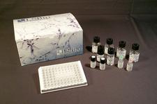
GDF15 ELISA Kit
Select
Sandwich
96-Well Strip Plate
Human
78 - 5000 pg/ml
Colorimetric - 450nm (TMB)
Plasma, Serum
1 Plate/$730
GDF15 ELISA Kit
Select
Sandwich
96-Well Strip Plate
Porcine
31.25 - 2000 pg/ml
Colorimetric - 450nm (TMB)
Plasma, Serum
1 Plate/$728
GDF15 Antibody - Western blot of recombinant GDF15.  This image was taken for the unconjugated form of this product. Other forms have not been tested.
Select
GDF15 Rabbit anti-Mouse Polyclonal (aa193-296) Antibody
Mouse
WB
Unconjugated
100 µl/$311; 200 µl/$355; 500 µl/$499; 1 ml/$571; 2 ml/$777; 5 ml/$1,261
GDF15 ELISA Kit
Select
Sandwich
96-Well Strip Plate
Human
15.6 - 1000 pg/ml
Colorimetric - 450nm (TMB)
Serum
1 Plate/$441
GDF15 ELISA Kit
Select
Sandwich
96-Well Strip Plate
Human
7.8 - 500 pg/ml
Colorimetric - 450nm (TMB)
Cell Culture Supernatants, Plasma, Serum
1 Plate/$590
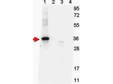
GDF15 Antibody - Anti-NAG-1 (H variant specific) Monoclonal Antibody - Western Blot. Western blot shows detection of recombinant NAG-1 protein present in Pichia pastoris whole cell lysates: lane 1 - yeast cell lysate expressing NAG-1 H variant with SUMO expression tag at 36 kD; lane 2 - yeast cell lysate expressing NAG-1 D variant with SUMO expression tag at 36 kD; lane 3 - yeast cell lysate expressing NAG-1 H variant; and lane 4 - yeast cell lysate expressing NAG-1 D variant. All lysates were run under reducing conditions. Primary antibody was used at a 1:1000 dilution in TBS containing 1% BSA and 0.2% Tween, and reacted overnight at 4°C. For detection, a 1:40000 dilution of peroxidase conjugated Gt-a-Mouse IgG secondary antibody (LS-C60680) was used in Blocking Buffer for Fluorescent Western Blot (MB-070) for 30 min at room temperature. Molecular weight estimation was made by comparison to prestained MW markers. Image was captured using the BioRad Versadoc 4000MP Imaging System.
Select
GDF15 Mouse anti-Human Monoclonal (7C12.B3.F2) Antibody
Human
ELISA, WB
Unconjugated
100 µg/$574
GDF15 Protein - Recombinant Growth Differentiation Factor 15 By SDS-PAGE
Select
E. coli
His, N-Terminal
14.1 kDa
10 µg/$288; 50 µg/$487; 100 µg/$780; 200 µg/$972; 1 mg/$2,109; 500 µg/$1,636; 5 mg/$3,403; 2 mg/$2,298
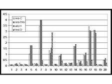
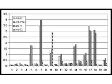
GDF15 Antibody - Anti-NAG-1 (D variant specific) Antibody - ELISA. In this sandwich ELISA, NAG-1 was captured from human serum using the following antibodies (see Related Products below): anti-NAG-1/GDF15 (C terminal specific), anti-NAG-1/GDF15 (N terminal specific (PAN)), anti-NAG-1/GDF15 (H-variant) and anti-NAG-1/GDF15 (D-variant) polyclonal antibodies. Micro titer plates were coated with capture antibody at 1 ug/mL. Control plates received PBS only (data not shown). After overnight incubation and blocking, independent experiments using 20 random normal human sera were performed. Neat normal sera were applied and incubated for 1 h at 37 °C. After washing, HRP conjugated anti-NAG-1/GDF15 (C terminal specific) antibody was added for detection at 100 ul per well at 1 ug/mL. Following further incubation for 1 hr at 37°C, the plates were washed and TMBE was added as an HRP substrate for 30 min. The reaction was stopped by 1 M H2SO4 and values were measured at 450nm.
Select
GDF15 Rabbit anti-Human Polyclonal Antibody
Human
ELISA, IHC, WB
Unconjugated
100 µg/$567
GDF15 Antibody - Anti-NAG-1/GDF15 (C-terminal) Antibody - Western Blot. Western blot of affinity purified anti-NAG-1/GDF15 (C-terminal) antibody shows detection NAG-1 purified from CHO cells as a 14 kD band corresponding to monomer and a 28 kD band corresponding to dimerized NAG-1. Samples were electrophoresed on a 4-20% gradient gel under reducing conditions. Lane 1 shows NAG-1 detection. Lane 2 shows reactivity is blocked when this antibody is pre-incubated with the immunizing peptide prior to Western blotting.
Select
GDF15 Rabbit anti-Human Polyclonal Antibody
Mouse, Human
ELISA, IHC, WB
Unconjugated
100 µg/$567
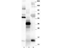
GDF15 Antibody - Anti-Mouse NAG-1/GDF15 Antibody - Western Blot. Western blot of affinity purified anti-mouse NAG-1/GDF15 antibody. The blot shows detection of recombinant MBP-NAG-1 fusion protein (60 kD) purified from E. coli (lane 1); yeast cell lysate expressing SUMO-mouse NAG-1 (42 kD) (lane 2), and R&D human NAG-1 monomer purified from CHO-K1 cells (14 kD) (lane 3). All lysates were run under reducing conditions. Primary antibody was used at a 1:1000 dilution in TBS containing 1% BSA and 0.2% Tween, and reacted overnight at 4C. Nag-1 was detected using a 1:40000 dilution of peroxidase conjugated Gt-a-Rabbit antibody (LS-C60865) in Blocking Buffer for Fluorescent Western Blot (MB-070) for 30 min at room temperature. Molecular weight estimation was made by comparison to prestained MW markers. Image was captured using the BioRad Versadoc 4000MP Imaging System. Other detection systems will yield similar results.
Select
GDF15 Rabbit anti-Mouse Polyclonal (C-Terminus) Antibody
Mouse, Human
ELISA, WB
Unconjugated
100 µg/$567
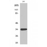
GDF15 Antibody - Western blot of GDF-15 antibody
Select
GDF15 Rabbit anti-Human Polyclonal (1-80 aa, Internal) Antibody
Human
ELISA, WB
Unconjugated
50 µg/$295; 100 µg/$335; 200 µg/$394
GDF15 Protein - Recombinant Growth Differentiation Factor 15 By SDS-PAGE
Select
E. coli
His, N-Terminal
34.9 kDa
10 µg/$289; 50 µg/$492; 200 µg/$982; 1 mg/$2,119; 5 mg/$3,431; 2 mg/$2,309
GDF15 ELISA Kit
Select
Sandwich
96-Well Strip Plate
Monkey
23.44 - 1500 pg/ml
Colorimetric - 450nm (TMB)
Plasma, Serum
1 Plate/$656
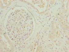
GDF15 Antibody - Immunohistochemistry of paraffin-embedded human colon cancer using GDF15 Antibody at diluton of 1:50
Select
GDF15 Rabbit anti-Human Polyclonal Antibody
Human
ELISA, IHC, WB
Unconjugated
50 µg/$294; 100 µg/$360
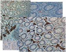
GDF15 Antibody - Immunohistochemistry of rabbit anti NAG1 antibody. Tissue: Human Colon at 20X in colon tissue at pH 6. Negative control of human colon tissue pH6 is shown in background.
Select
GDF15 Rabbit anti-Human Polyclonal Antibody
Mouse, Human
ELISA, IHC, WB
Unconjugated
25 µl/$304
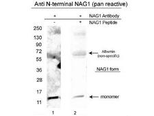
GDF15 Antibody - Anti-NAG-1/GDF15 (N-terminal specific) Antibody - Western Blot. Western blot of affinity purified anti-NAG-1/GDF15 (N-terminal specific) antibody shows detection of a 14 kD band corresponding to recombinant human NAG-1 purified from CHO cells. Samples were electrophoresed on a 4-20% gradient gel under reducing conditions. Lane 1 shows NAG-1 detection. Lane 2 shows reactivity is greatly diminished when this antibody is preincubated with the immunizing peptide prior to Western blotting.
Select
GDF15 Rabbit anti-Human Polyclonal Antibody
Human
ELISA, IHC, WB
Unconjugated
100 µg/$567
GDF15 Antibody - Anti-NAG-1 (H variant specific) Antibody - ELISA. In this sandwich ELISA, NAG-1 was captured from human serum using the following antibodies (see Related Products below): anti-NAG-1/GDF15 (C terminal specific), anti-NAG-1/GDF15 (N terminal specific (PAN)), anti-NAG-1/GDF15 (H-variant) and anti-NAG-1/GDF15 (D-variant) polyclonal antibodies. Micro titer plates were coated with capture antibody at 1 ug/mL. Control plates received PBS only (data not shown). After overnight incubation and blocking, independent experiments using 20 random normal human sera were performed. Neat normal sera were applied and incubated for 1 h at 37 °C. After washing, HRP conjugated anti-NAG-1/GDF15 (C terminal specific) antibody was added for detection at 100 ul per well at 1 ug/mL. Following further incubation for 1 hr at 37°C, the plates were washed and TMBE was added as an HRP substrate for 30 min. The reaction was stopped by 1 M H2SO4 and values were measured at 450nm.
Select
GDF15 Rabbit anti-Human Polyclonal Antibody
Human
ELISA, IHC, WB
Unconjugated
100 µg/$567
Viewing 1-25 of 147 product results


If you do not find the reagent or information you require, please contact Customer.Support@LSBio.com to inquire about additional products in development.

PLEASE NOTE

For RESEARCH USE ONLY. Intended for use by laboratory professionals. Not intended for human diagnostic or therapeutic purposes.

The data on this page has been compiled from LifeSpan internal sources, the National Center for Biotechnology Information (NCBI), and The Universal Protein Resource (UniProt).